When neuroscientist Jakob Seidlitz took his 15-month-old son to the paediatrician for a check-up last week, he left feeling unsatisfied. There wasn’t anything wrong with his son — the youngster seemed to be developing at a typical pace, according to the height and weight charts the physician used. What Seidlitz felt was missing was an equivalent metric to gauge how his son’s brain was growing. “It is shocking how little biological information doctors have about this critical organ,” says Seidlitz, who is based at the University of Pennsylvania in Philadelphia.
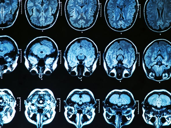
Soon, he might be able to change that. Working with colleagues, Seidlitz has amassed more than 120,000 brain scans — the largest collection of its kind — to create the first comprehensive growth charts for brain development. The charts show visually how human brains expand quickly early in life and then shrink slowly with age. The sheer magnitude of the study, published in Nature on 6 April, has stunned neuroscientists, who have long had to contend with reproducibility issues in their research, in part because of small sample sizes. Magnetic resonance imaging (MRI) is expensive, meaning that scientists are often limited in the number of participants they can enrol in experiments.
“The massive data set they assembled is extremely impressive and really sets a new standard for the field,” says Angela Laird, a cognitive neuroscientist at Florida International University in Miami.
Even so, the authors caution that their database isn’t completely inclusive — they struggled to gather brain scans from all regions of the globe. The resulting charts, they say, are therefore just a first draft, and further tweaks would be needed to deploy them in clinical settings.
If the charts are eventually rolled out to paediatricians, great care will be needed to ensure that they are not misinterpreted, says Hannah Tully, a paediatric neurologist at the University of Washington in Seattle. “A big brain is not necessarily a well-functioning brain,” she says.
No easy task
Because brain structure varies significantly from person to person, the researchers had to aggregate a huge number of scans to create an authoritative set of growth charts with statistical significance. That’s no easy task, says Richard Bethlehem, a neuroscientist at the University of Cambridge, UK, and a co-author of the study. Instead of running thousands of scans themselves, which would take decades and be prohibitively costly, the researchers turned to already-completed neuroimaging studies.
Bethlehem and Seidlitz sent e-mails to researchers all over the world asking if they would share their neuroimaging data for the project. The duo was amazed by the number of replies, which they attribute to the COVID-19 pandemic giving researchers less time in their laboratories and more time than usual with their e-mail inboxes.
In total, the team aggregated 123,894 MRI scans from 101,457 people, who ran the gamut from fetuses 16 weeks after conception to 100-year-old adults. The scans included brains from neurotypical people, as well as people with a variety of medical conditions, such as Alzheimer’s disease, and neurocognitive differences, including autism spectrum disorder. The researchers used statistical models to extract information from the images, and ensure that the scans were directly comparable, no matter what type of MRI machine had been used.
.jpeg)
The end result is a set of charts plotting several key brain metrics by age. Some metrics, such as grey-matter volume and mean cortical thickness (the width of the grey matter) peak early in a person’s development, whereas the volume of white matter (found deeper in the brain) tends to peak by around age 30 (see ‘Brain change’). The data on ventricular volume (the amount of cerebrospinal fluid in the brain), in particular, surprised Bethlehem. Scientists knew that this volume increases with age, because it is typically associated with brain atrophy, but Bethlehem was shocked by how rapidly it tends to grow in late adulthood.
A first draft
The study comes on the heels of a bombshell paper published in Nature on 16 March showing that most brain-imaging experiments contain too few scans to reliably detect links between brain function and behaviour, meaning that their conclusions might be incorrect. Given this finding, Laird expects the field to move towards adopting a framework similar to the one used by Seidlitz and Bethlehem, to increase statistical power.
To amass so many data sets is akin to a “diplomatic masterpiece”, says Nico Dosenbach, a neuroscientist at Washington University in St. Louis, Missouri, who co-authored the 16 March study. He says this is the scale on which researchers should operate when aggregating brain images.
Despite the size of the data set, Seidlitz, Bethlehem and their colleagues acknowledge that their study suffers from a problem endemic to neuroimaging studies — a remarkable lack of diversity. The brain scans they collected come mainly from North America and Europe, and disproportionately reflect populations that are white, university-aged, urban and affluent. This limits the generalizability of the findings, says Sarah-Jayne Blakemore, a cognitive neuroscientist at the University of Cambridge. The study includes only three data sets from South America and one from Africa — accounting for around 1% of all the brain scans used in the study.
Billions of people worldwide lack access to MRI machines, making diverse brain-imaging data difficult to come by, Laird says. But the authors haven’t stopped trying. They have launched a website where they intend to update their growth charts in real time as they receive more brain scans.
With big data sets, big responsibility
Another challenge was determining how to give proper credit to the owners of the brain scans used to construct the charts. Some of the scans came from open-access data sets, but others were closed to researchers. Most of the closed-data scans hadn’t yet been processed in a way that would allow them to be incorporated into the growth charts, so their owners did extra work to share them. These scientists were then named as authors of the paper.
Meanwhile, the owners of the open data sets received only a citation in the paper — which doesn’t hold as much prestige for researchers seeking funding, collaborations and promotions. Seidlitz, Bethlehem and their colleagues processed these data. In most cases, Bethlehem says that there was essentially no direct contact with the owners of these data sets. The paper lists about 200 authors and cites the work of hundreds of others who contributed brain scans.
There are a number of reasons that data sets might be closed: for instance, to protect the privacy of health data, or because researchers don’t have the resources to make them public. But this doesn’t make it fair that the researchers who opened their data sets didn’t get authorship, the authors say. In their paper’s Supplementary Information, they argue that the situation “perversely disincentivises open science, since the people who do most to make their data openly available could be least likely to merit recognition”. Bethlehem and Seidlitz contend that authorship guidelines from journals, including Nature — which say that each author is expected to have made “substantial contributions” to, for example, the analysis or interpretation of data — are an obstacle. (Nature’s news team is editorially independent of its publisher.)
A Nature spokesperson responds that the issue was “considered carefully by the editors and authors according to our authorship policies” and that “all datasets were appropriately credited per our data citation policy”.
Ultimately, these concerns can be traced back to how researchers are evaluated by the scientific enterprise, says Kaja LeWinn, a social epidemiologist at the University of California, San Francisco, who studies neurodevelopment. She says that it’s incumbent on all of the relevant stakeholders — including funders, journals and research institutions — to re-evaluate how brain science can be properly recognized and rewarded, especially as these types of large-scale study become more common.
This article is reproduced with permission and was first published on April 6 2022.


