In 1968 an exhibit entitled Cybernetic Serendipity: The Computer and the Arts was held at the Institute of Contemporary Arts in London. The first major event of its kind, Cybernetic Serendipity’s aim was to “present an area of activity which manifests artists’ involvement with science, and the scientists’ involvement with the arts,” wrote British art critic Jasia Reichardt, who curated the exhibit. Even though it was an art show, “most of the participants in the exhibition were scientists,” Reichardt said in a 2014 video. “Artists didn’t have computers in the 1960s.” A lot has changed since then, however. Computers, no longer the commodity of a select few, help artists to deviate from more traditional mediums.
The changes since the 1960s are well-reflected in the entries for the 2020 Art of Neuroscience competition, held by the Netherlands Institute for Neuroscience. Now marking its 10th year, the contest features some highly technological pieces and others grounded in classical methods, such as drawing with pen on paper. The winning entries were created by independent artists, as well as working scientists, demonstrating that art and neuroscience can inspire both professions. A winner and four honorable mentions were selected from dozens of submitted works. And seven pieces were chosen by Scientific American as Editors’ Picks. (Photography editor Liz Tormes served on the panel of judges for the competition.)
WINNER
31-3594
by Lidija Kononenko
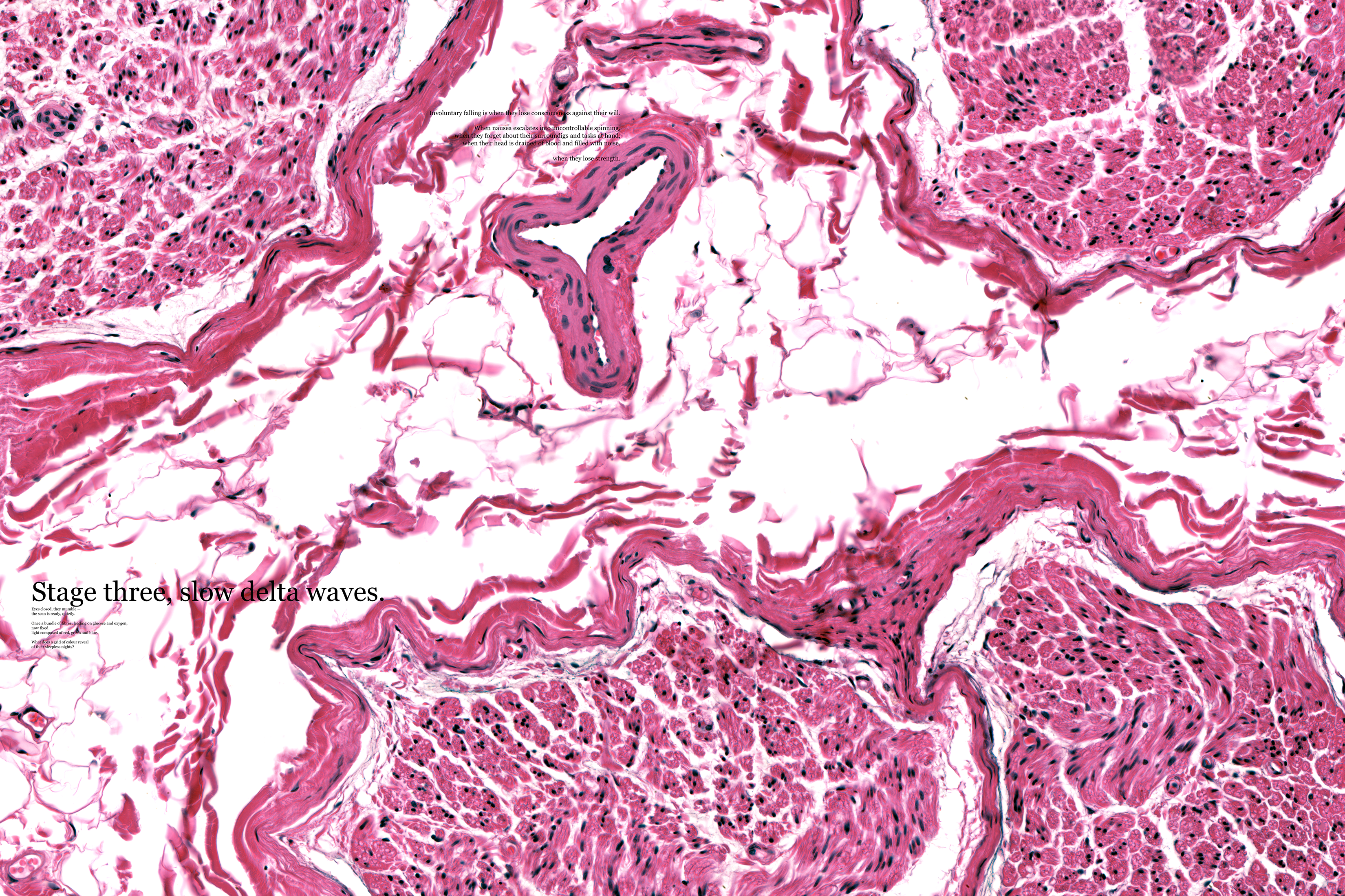
Artist Kononenko described this interactive piece as “a microscope specimen, a map of symptoms, and an investigation of the unknown” in a statement accompanying it. Viewers can zoom in and explore the details of a microscope image of the peripheral nerve system, which is overlaid by textual facts and poetic phrases about sleep.
Sleep is “a voluntary act of losing one’s own consciousness,” Kononenko explained in her statement. The poetic snippets resemble the fragmented thoughts humans have while falling asleep. And zooming in and out of the image represents the transition between wakefulness and sleep. Additionally, 31-3594 allows the viewer to act as a pathologist, achieving the goal of blending neuroscience and art. In assessing this unique piece, the jurors praised it for “the interactivity and playful combination of imagery of a human peripheral nerve with a text-based story that unfolds at various scales and highlights the role of the nervous system in the human condition.”
HONORABLE MENTION
Red Haze
by Nicki Coveña
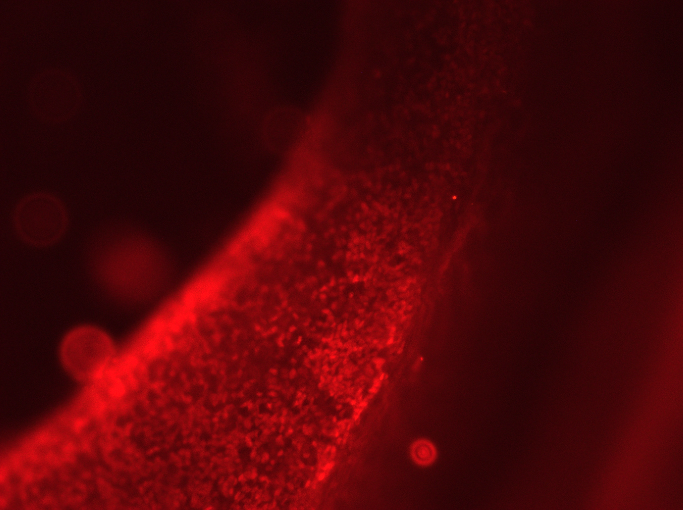
A tsunami of red dots dominates this image by neuroscientist Coveña. The bright red color comes from a fluorescent protein, which was used to visualize the workings of TBR1—a gene that synthesizes the protein that regulates the information transfer from DNA to messenger RNA in vertebrate embryo development. “The out-of-focus view makes one guess at what details are hidden below,” the jurors wrote.
HONORABLE MENTION
Bdl
by Paméla Simard
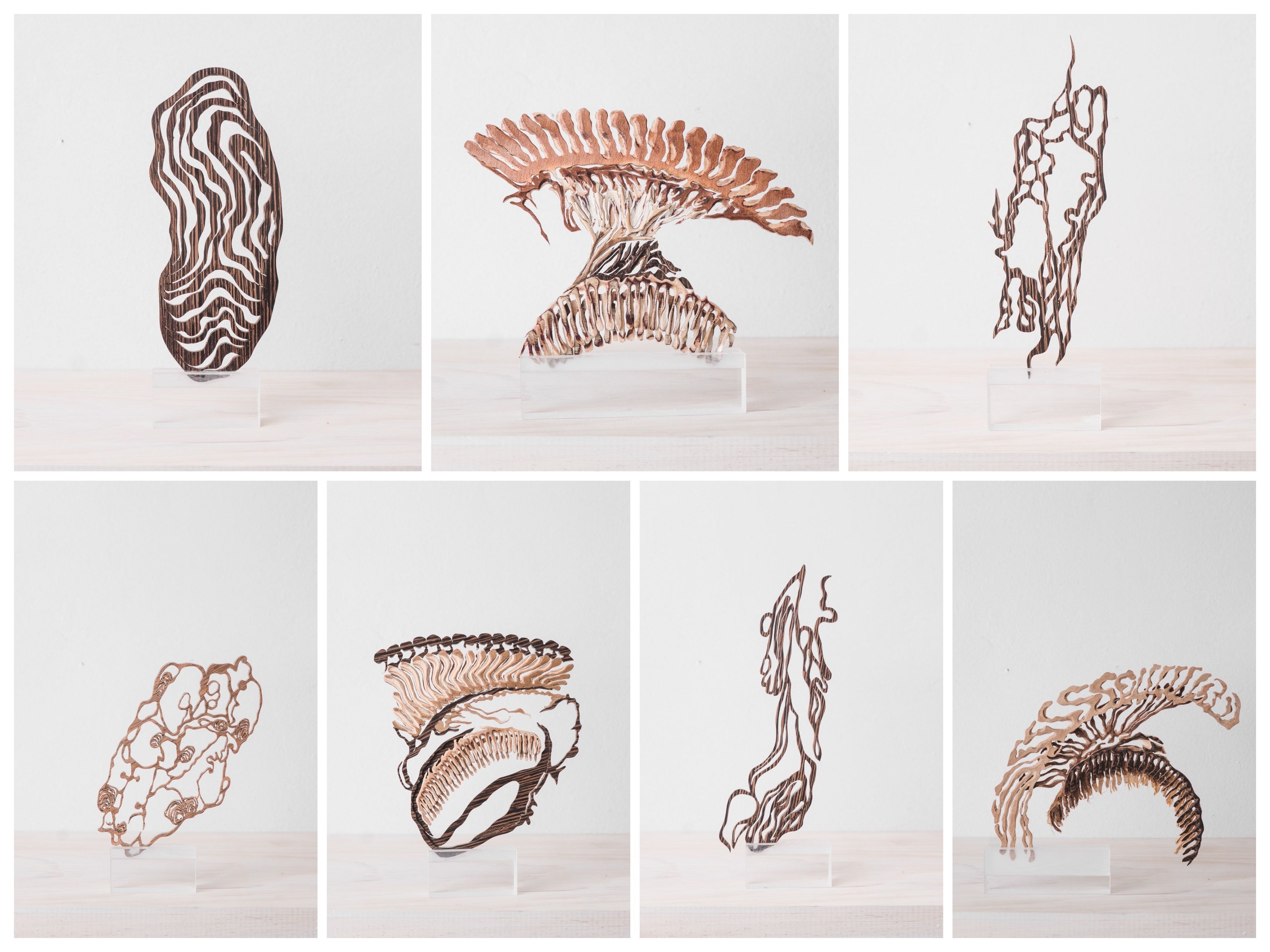
Artist Simard partnered with Hunter Shaw, a neuroscientist then at McGill University, to create a series of delicate wooden sculptures. “The various installations were created from fluorescent microscopy images representing the visual system of the fruit fly brain,” Simard wrote in her statement. The intricate details of the fruit fly visual system were made possible by first laminating the thin slices of different types of wood together, then hand cutting the result to mimic the microscope images.
HONORABLE MENTION
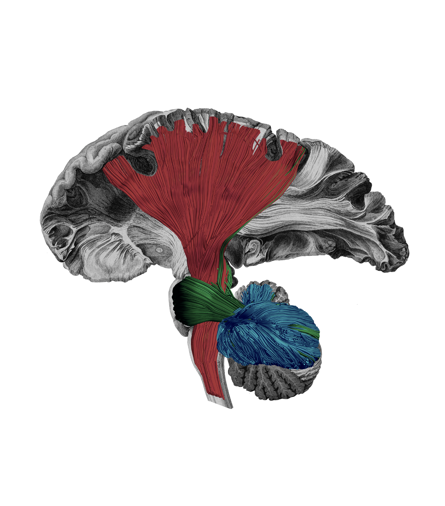
Motor White Matter Networks of the Human Brain
by Sanja Budisavljevic
In this piece, neuroscientist Budisavljevic superimposed color onto a 19th-century black-and-white drawing of a brain based on a postmortem dissection. Each color indicates a different “highway,” or white matter pathway connecting particular regions of gray matter and allowing information to be transferred. Red indicates the most prominent highway, which links the cortex and spinal cord. “This pathway carries the messages to and from the body and allows us to function in our sensory world,” Budisavljevic says. Green represents the connection that supports coordination, and blue shows the one that regulates movements.
HONORABLE MENTION
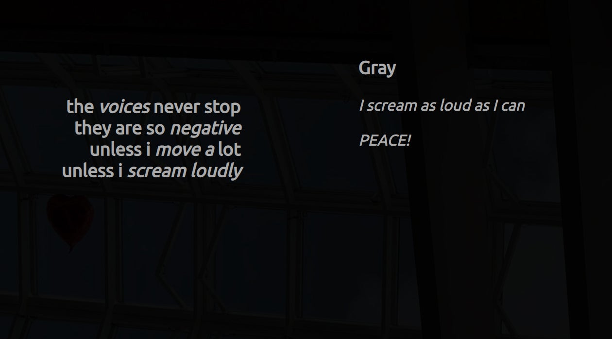
Psychoetry
by Frank Gerritse and Janna de Boer
This interactive piece, created by psychiatrists Gerritse and de Boer, allows viewers to experience auditory verbal hallucinations—voices that appear in one’s head. “People who hear voices often feel trapped and experience little or no control,” Gerritse and de Boer wrote in a brochure about the project. Singing, screaming, or getting up and moving around, however, help some of them to perceive the voices as less negative. The researchers programmed their piece to emulate this experience, as well as to allow “people to influence both the amount and the content of the hallucinations,” they wrote. When the interactive launches, users face disparaging text that simulates negative self-talk; this material was recorded and transcribed from real hallucinations. They can also hear loud noises by clicking a headphone icon. To make the text display stop for a few seconds, viewers can yell at their computer after enabling its microphone access. Similarly, when they jiggle their mouse or quickly move their fingers across their trackpad—imitating the real-life movements that help disrupt hallucinations—the text becomes “nicer” and less critical.
Editor’s Note: Viewer discretion is advised, because some of the text includes obscene language. The Web site works best with Chrome, and the browser’s auto-translate function is recommended for non-Dutch speakers.
EDITOR’S PICK
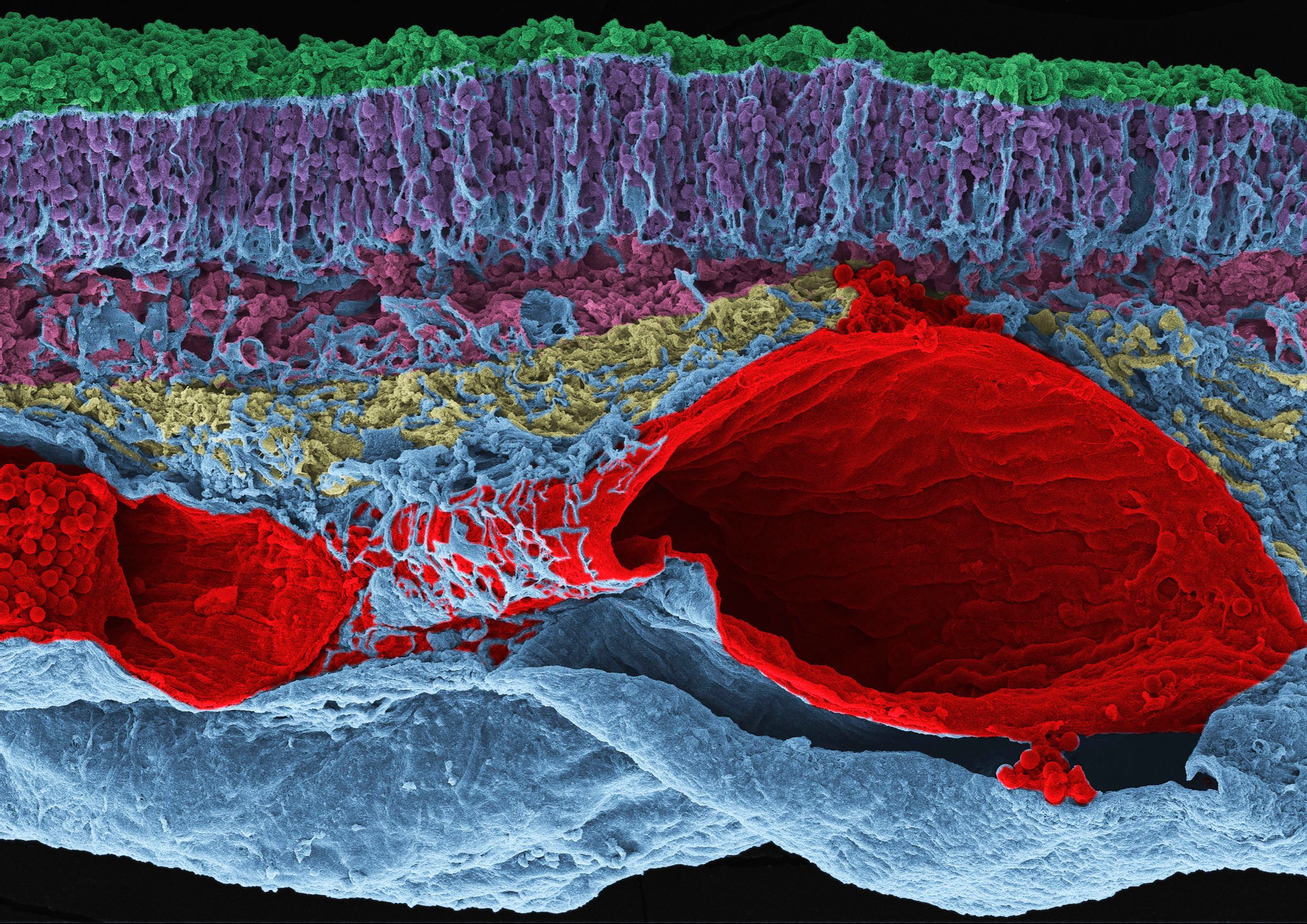
Whale Retina Rainbow
by Elena Vecino Cordero and Luis López Vecino
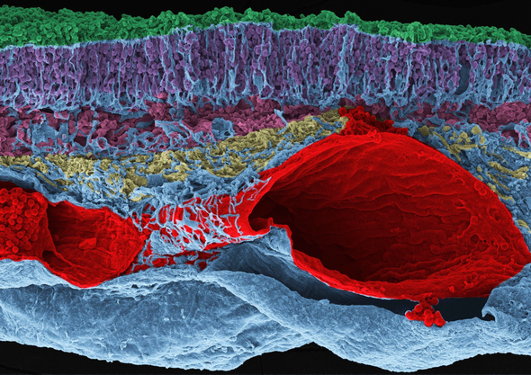
In February 2019 the death of a whale in Sopelana Beach in Spain made the local news. The beach happened to be close to the University of the Basque Country, where biologist Vecino Cordero works. Seizing the opportunity, she and some volunteers extracted the eye of the whale and took it back to her ophthalmology research group for further study. The image was produced as a part of their research. The whale’s retina was imaged using scanning electron microscopy. And later López Vecino added the colors using Adobe Photoshop.
EDITOR’S PICK
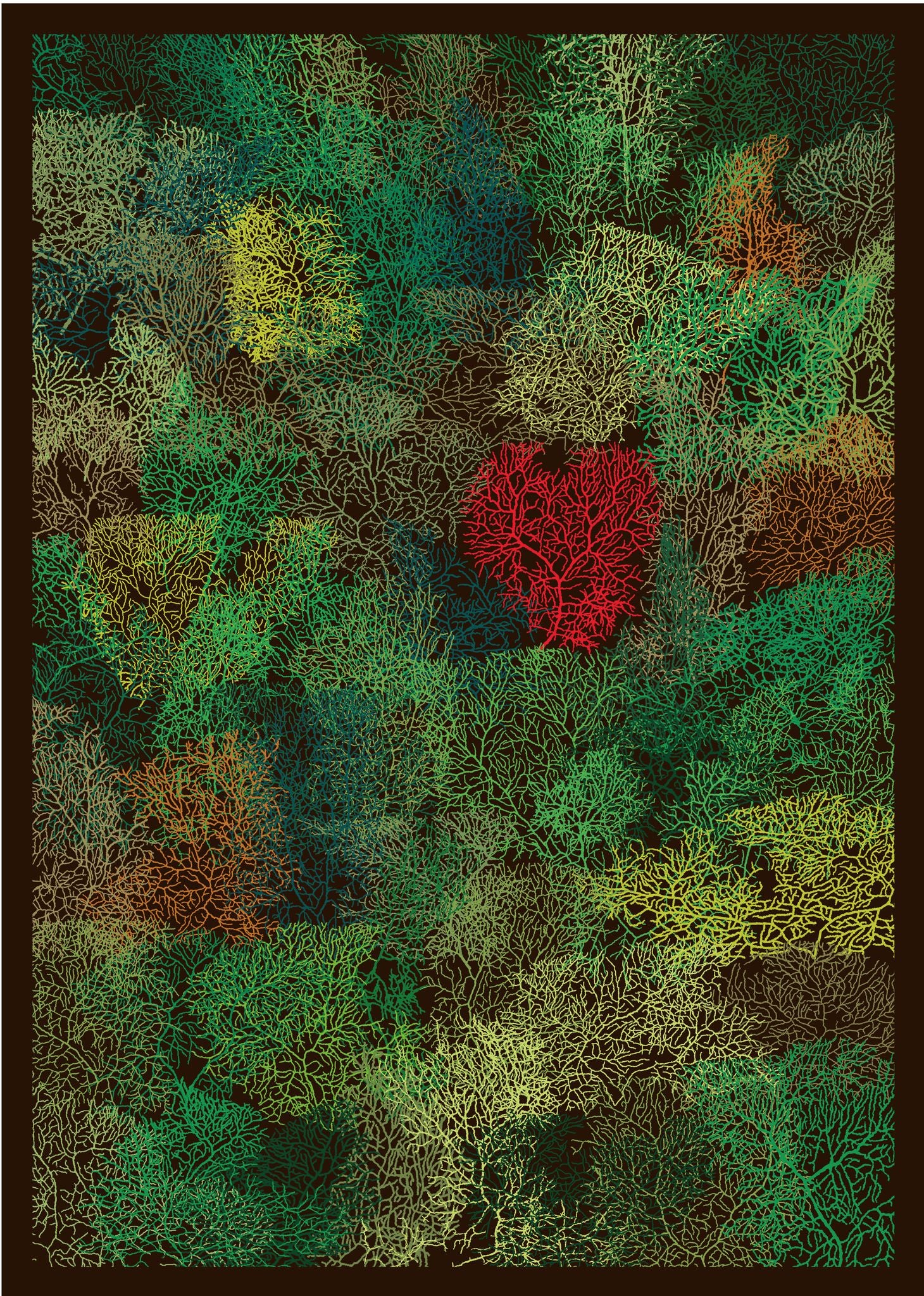
The Protection of Nature Starts in Our Mind
by Robert Luck
Luck is a neuroscientist at Heidelberg University in Germany who studies the development of the cerebellum, located where the spinal cord meets the brain. Alarmed by climate change and deforestation, he created a “mind forest” that resembles bird’s-eye-view photographs of real forests. The “trees” are 65 individually traced images of mice’s Purkinje neurons, which play important roles in controlling coordination and movements. “I chose the number 65 to represent the number of years needed for the rainforest to regrow and gain back at least 80% of its diversity,” Luck wrote in his statement. “[Sixty-five] years—a human lifetime!”
EDITOR’S PICK
Shelter in Place
by Geinene Carson
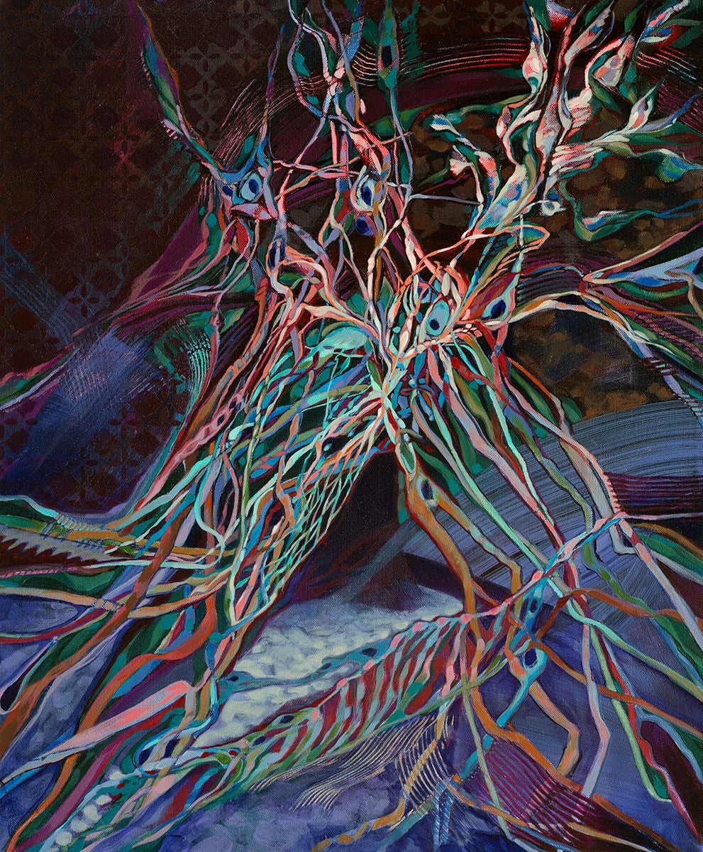
As its title suggests, this piece represents “the artist’s interpretation of the pandemic experience” while sheltering in place because of COVID-19, according to artist Carson’s statement. This acrylic-on-canvas piece is a part of a series entitled Neuron, which started as “visual prayers for our daughter with a rare genetic disorder,” Carson wrote on her Web site. While Shelter in Place implies physical restrictions, Carson, who is based in Atlanta, draws inspiration from the neural network, “because as important as our physical surroundings are to our state of living, our thought life holds the key to thriving within whatever the circumstances may be,” she wrote.
EDITOR’S PICK
Sensing Spin
by Dan Jagger
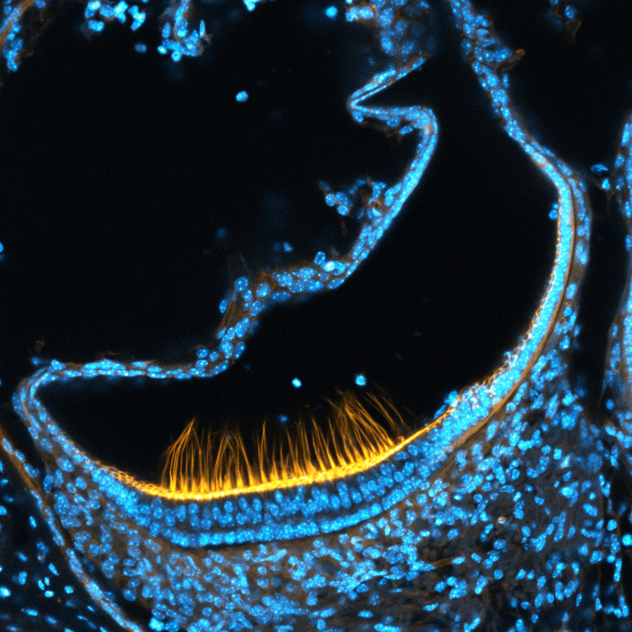
Physiologist Jagger used a high-resolution microscope to capture this image. It shows mechanosensory hair cells located in the inner ear that play a role in the sense of balance. A protein called actin is within bundles of stereocilia and is stained yellow. Actin helps the bundles to stand upright, so when the human head turns, they can detect the movement of the fluid they are immersed in. The hair-cell nuclei are stained with cyan.
EDITOR’S PICK
Memories and Patterns: Oligodendrocytes
by Shanthi Chandrasekar
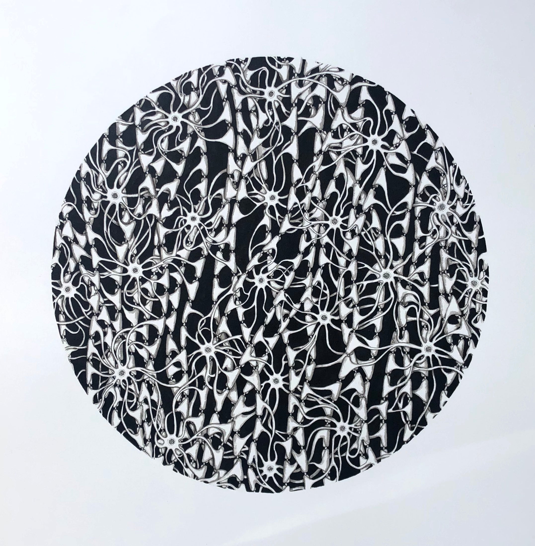
Oligodendrocytes are glial cells that support and insulate long neuronal axons. The cells’ lipid membrane wraps around the axons to strengthen the structure, as well as to help neurons to send signals quickly. “A single oligodendrocyte can connect with multiple axons,” artist Chandrasekar wrote in her statement. “In this [pen-and-ink] drawing, I have tried to bring out the connectedness of the oligodendrocytes and the axons.”
EDITOR’S PICK
Bridges between Genesis and Neuroscience: Triplets
by Rui Rodrigues
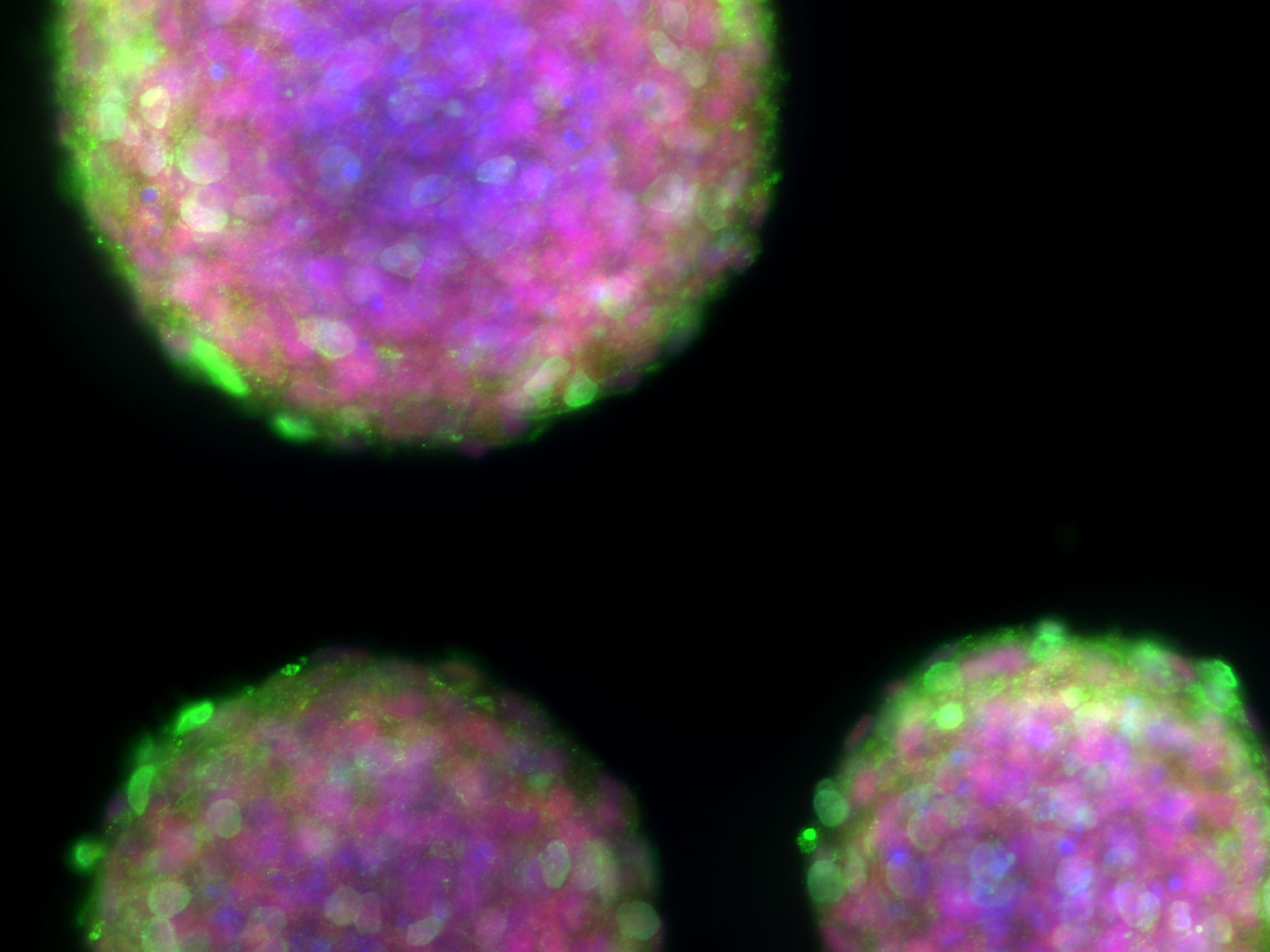
This image features three neurospheres—clusters of neural stem or progenitor cells—that are similar in size and shape. Because of their similarity, neurobiologist Rodrigues entitled the piece Triplets. The vibrant colors come from “antibodies coupled with fluorescent tags to label specific proteins,” he says.
EDITOR’S PICK
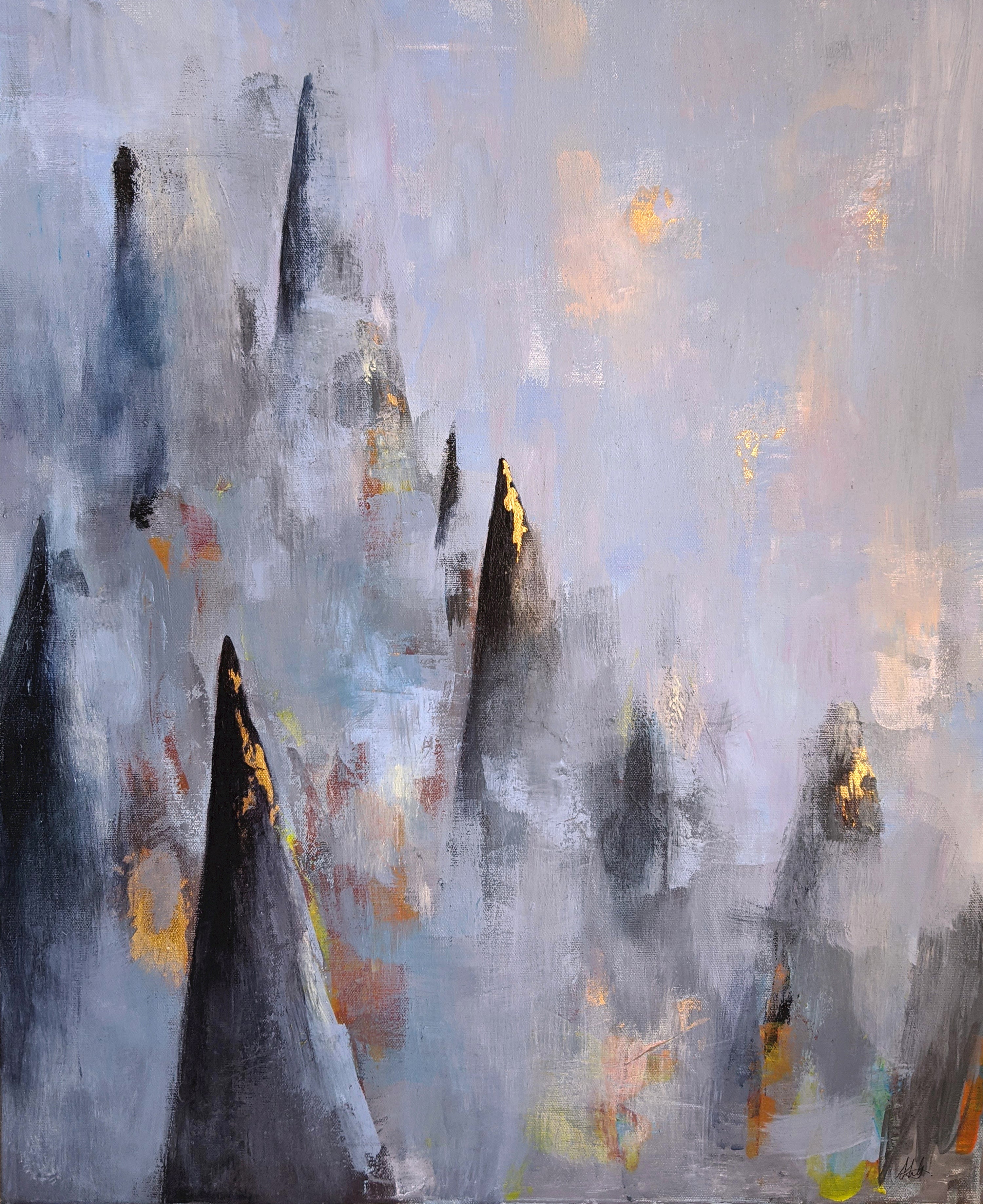
Fading Engram
by Alexandra Leighton
“Our experience of the world and our sense of self are shaped by what we remember,” neuroscientist Leighton wrote in her statement. “Some memories stay crystal clear, others become distorted or slip away as time passes by.” Well-defined and blurred peaks each represent sharp and faded memories. This piece—made with acrylic and gold leaf on canvas—was inspired by images of hippocampal neurons, which are known to play a significant role in memory formation.
